Long Bone Labeled Endosteum / Structure Of Long Bone Animal Systems - Endochondrial bone formation begins with a cartilage precursor that proliferates, hypertrophies, and becomes imbedded in the core of bone.
Long Bone Labeled Endosteum / Structure Of Long Bone Animal Systems - Endochondrial bone formation begins with a cartilage precursor that proliferates, hypertrophies, and becomes imbedded in the core of bone.. Microscopic bone structure compact bone is organized as parallel columns, known as haversian systems, which run lengthwise down the axis of long bones. The diaphysis is the main or midsection (shaft) of a long bone.it is made up of cortical bone and usually contains bone marrow and adipose tissue (fat). Endochondrial bone formation begins with a cartilage precursor that proliferates, hypertrophies, and becomes imbedded in the core of bone. Located in the main shaft of a long bone (consisting mostly of compact bone), the medullary cavity has walls composed of spongy bone (cancellous bone) and is lined with a thin, vascular membrane. Rather, bone is deposited on the surface of calcified.
The cavernous sinuses are paired dural venous sinuses. The inside of the diaphysis, at the border between the cortical and cancellous bone and lining the trabeculae, is lined by endosteum. Sep 07, 2017 · endosteum. Microscopic bone structure compact bone is organized as parallel columns, known as haversian systems, which run lengthwise down the axis of long bones. Rather, bone is deposited on the surface of calcified.
Print A P Chapter 6 Bones And Skeletal Tissues Flashcards Easy Notecards from www.easynotecards.com A small mineralized spicule that forms a network in spongy bone; The inside of the diaphysis, at the border between the cortical and cancellous bone and lining the trabeculae, is lined by endosteum. An important point, here, is that cartilage does not become bone. The endosteum greatly resembles the periosteum, consisting of a thin layer of very tough fibrous tissue, which also contains nerve cells. Endochondrial bone formation begins with a cartilage precursor that proliferates, hypertrophies, and becomes imbedded in the core of bone. Spongy bone is prominent in regions where the bone is less dense and at the ends of long bones where the bone has to be more compressible due to stresses that arrive from many directions. The cavernous sinuses are paired dural venous sinuses. Microscopic bone structure compact bone is organized as parallel columns, known as haversian systems, which run lengthwise down the axis of long bones.
The inside of the diaphysis, at the border between the cortical and cancellous bone and lining the trabeculae, is lined by endosteum.
Rather, bone is deposited on the surface of calcified. The inside of the diaphysis, at the border between the cortical and cancellous bone and lining the trabeculae, is lined by endosteum. Sep 07, 2017 · endosteum. However, the medullary cavity is the area inside any bone (long, flat, etc.) that holds the bone marrow. The cavernous sinuses are paired dural venous sinuses. The endosteum greatly resembles the periosteum, consisting of a thin layer of very tough fibrous tissue, which also contains nerve cells. A small mineralized spicule that forms a network in spongy bone; It is a middle tubular part composed of compact bone which surrounds a central marrow cavity which contains red or yellow marrow. Spongy bone is prominent in regions where the bone is less dense and at the ends of long bones where the bone has to be more compressible due to stresses that arrive from many directions. The rounded end of any long bone Microscopic bone structure compact bone is organized as parallel columns, known as haversian systems, which run lengthwise down the axis of long bones. Located in the main shaft of a long bone (consisting mostly of compact bone), the medullary cavity has walls composed of spongy bone (cancellous bone) and is lined with a thin, vascular membrane. An important point, here, is that cartilage does not become bone.
Rather, bone is deposited on the surface of calcified. Located in the main shaft of a long bone (consisting mostly of compact bone), the medullary cavity has walls composed of spongy bone (cancellous bone) and is lined with a thin, vascular membrane. The cavernous sinuses are paired dural venous sinuses. Endochondrial bone formation begins with a cartilage precursor that proliferates, hypertrophies, and becomes imbedded in the core of bone. The diaphysis is the main or midsection (shaft) of a long bone.it is made up of cortical bone and usually contains bone marrow and adipose tissue (fat).
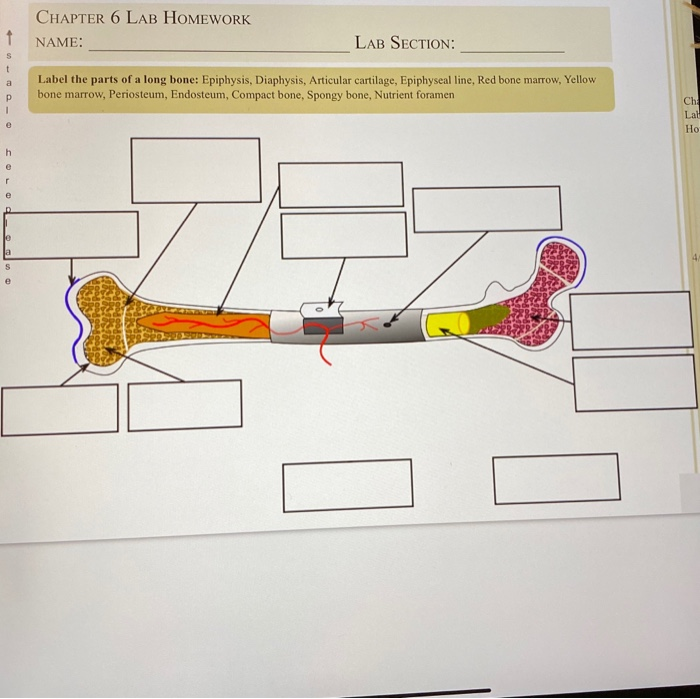
Chapter 6 Lab Homework Name 1 Lab Section S T A Chegg Com from media.cheggcdn.com Spongy bone is prominent in regions where the bone is less dense and at the ends of long bones where the bone has to be more compressible due to stresses that arrive from many directions. Microscopic bone structure compact bone is organized as parallel columns, known as haversian systems, which run lengthwise down the axis of long bones. Sep 07, 2017 · endosteum. Located in the main shaft of a long bone (consisting mostly of compact bone), the medullary cavity has walls composed of spongy bone (cancellous bone) and is lined with a thin, vascular membrane. Gross anatomy the cavernous sinus is located on either side of the pituitary fossa and body of the sphenoid bone between the endosteal and meningeal layers of the dura. The diaphysis is the main or midsection (shaft) of a long bone.it is made up of cortical bone and usually contains bone marrow and adipose tissue (fat). However, the medullary cavity is the area inside any bone (long, flat, etc.) that holds the bone marrow. An important point, here, is that cartilage does not become bone.
The endosteum greatly resembles the periosteum, consisting of a thin layer of very tough fibrous tissue, which also contains nerve cells.
The endosteum greatly resembles the periosteum, consisting of a thin layer of very tough fibrous tissue, which also contains nerve cells. A small mineralized spicule that forms a network in spongy bone; This method is most common in long bones of the body (like the shaft of the femur). An important point, here, is that cartilage does not become bone. The rounded end of any long bone Gross anatomy the cavernous sinus is located on either side of the pituitary fossa and body of the sphenoid bone between the endosteal and meningeal layers of the dura. It is a middle tubular part composed of compact bone which surrounds a central marrow cavity which contains red or yellow marrow. However, the medullary cavity is the area inside any bone (long, flat, etc.) that holds the bone marrow. Rather, bone is deposited on the surface of calcified. The cavernous sinuses are paired dural venous sinuses. The diaphysis is the main or midsection (shaft) of a long bone.it is made up of cortical bone and usually contains bone marrow and adipose tissue (fat). Endochondrial bone formation begins with a cartilage precursor that proliferates, hypertrophies, and becomes imbedded in the core of bone. Located in the main shaft of a long bone (consisting mostly of compact bone), the medullary cavity has walls composed of spongy bone (cancellous bone) and is lined with a thin, vascular membrane.
An important point, here, is that cartilage does not become bone. Spongy bone is prominent in regions where the bone is less dense and at the ends of long bones where the bone has to be more compressible due to stresses that arrive from many directions. The diaphysis is the main or midsection (shaft) of a long bone.it is made up of cortical bone and usually contains bone marrow and adipose tissue (fat). However, the medullary cavity is the area inside any bone (long, flat, etc.) that holds the bone marrow. The rounded end of any long bone
Unit 4 Standard 2 Quizlet Flashcards Quizlet from o.quizlet.com The inside of the diaphysis, at the border between the cortical and cancellous bone and lining the trabeculae, is lined by endosteum. Microscopic bone structure compact bone is organized as parallel columns, known as haversian systems, which run lengthwise down the axis of long bones. Gross anatomy the cavernous sinus is located on either side of the pituitary fossa and body of the sphenoid bone between the endosteal and meningeal layers of the dura. Endochondrial bone formation begins with a cartilage precursor that proliferates, hypertrophies, and becomes imbedded in the core of bone. The cavernous sinuses are paired dural venous sinuses. This method is most common in long bones of the body (like the shaft of the femur). A small mineralized spicule that forms a network in spongy bone; An important point, here, is that cartilage does not become bone.
Located in the main shaft of a long bone (consisting mostly of compact bone), the medullary cavity has walls composed of spongy bone (cancellous bone) and is lined with a thin, vascular membrane.
The inside of the diaphysis, at the border between the cortical and cancellous bone and lining the trabeculae, is lined by endosteum. The cavernous sinuses are paired dural venous sinuses. This method is most common in long bones of the body (like the shaft of the femur). An important point, here, is that cartilage does not become bone. Sep 07, 2017 · endosteum. It is a middle tubular part composed of compact bone which surrounds a central marrow cavity which contains red or yellow marrow. A small mineralized spicule that forms a network in spongy bone; Microscopic bone structure compact bone is organized as parallel columns, known as haversian systems, which run lengthwise down the axis of long bones. The endosteum greatly resembles the periosteum, consisting of a thin layer of very tough fibrous tissue, which also contains nerve cells. Spongy bone is prominent in regions where the bone is less dense and at the ends of long bones where the bone has to be more compressible due to stresses that arrive from many directions. The rounded end of any long bone Gross anatomy the cavernous sinus is located on either side of the pituitary fossa and body of the sphenoid bone between the endosteal and meningeal layers of the dura. The diaphysis is the main or midsection (shaft) of a long bone.it is made up of cortical bone and usually contains bone marrow and adipose tissue (fat).


Posting Komentar
0 Komentar